Advanced Light Microscopy
Our advanced light microscopy facility is home to:
- A state-of-the-art fully automated stereozoom microscope (the Zeiss AxioZoom V16)
- Two scanning-laser confocal microscopes with incubation chambers and autofocus (Zeiss LSM80 with Airyscan SR module; Leica Stellaris 5 with resonant scanner, super-Z Galvo stage, TauSense and lightning SR modes)
- One spinning-disk confocal microscope with incubation chamber (Crest X-lightv3 with optosplit and dual cameras)
- A deconvolution fluorescence imaging platform (DeltaVision2)
- Several widefield epifluorescence systems
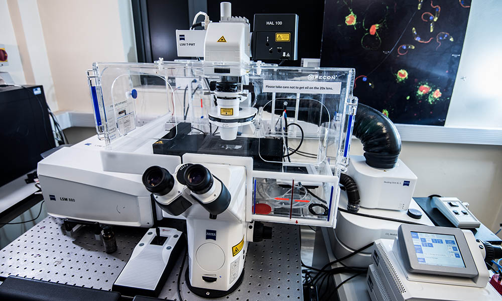
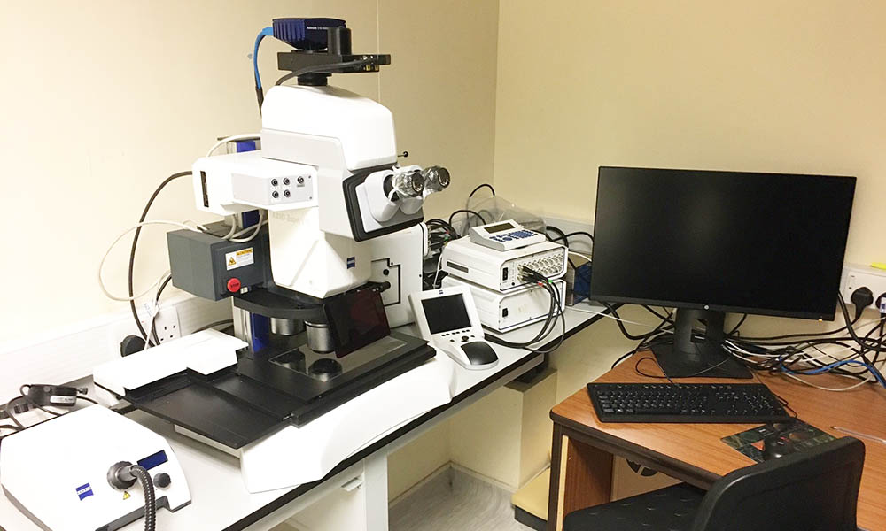
Zeiss LSM 880 with Airyscan Confocal Microscope
Zeiss LSM 880 confocal microscope with Airyscan module for near super-resolution imaging. This fully automated microscope is used for optical sectioning, tiling and time lapse experiments for samples on microscope slides, petri dishes or multi-well plates. The system is fully enclosed in an environmental chamber allowing control of temperature and CO2 gas delivery during live-cell imaging. The Airyscan module opens up further applications such as imaging of individually labelled molecules or viral infection of cells while also allowing improved resolution for live-cell imaging without increased phototoxicity or photo-bleaching.
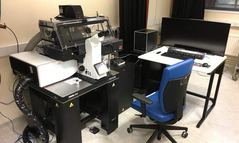
Leica Stellaris 5 Confocal Microscope
Leica Stellaris 5 confocal microscope with integrated white light laser (WLL), and Power HyD S silicon detectors. The fully tuneable WLL allows tuning of excitation between 485nm and 695nm with 1nm precision, while imaging up to 8 laser lines with freely configurable detection up to 850nm. The Power HyD detectors enable TauSense Technology, giving access to fluorescence lifetime-based information and intensity data simultaneously. The setup also includes Lightening SR mode, an information extraction technology that provides near super resolution images in real-time. A Super-Z Galvo stage affords fast, accurate Z-scanning and vibration free imaging. The system provides full environmental control for live imaging.
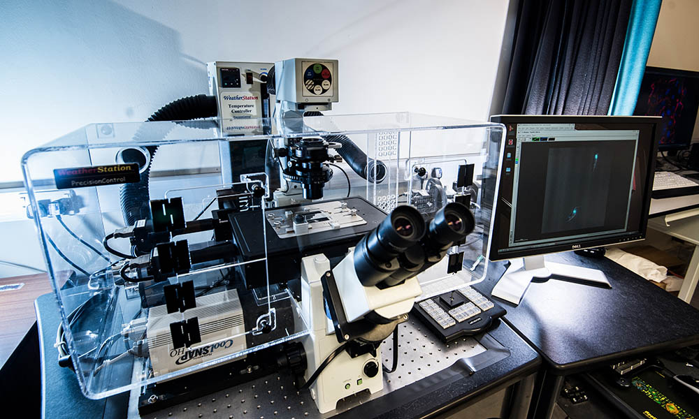
Crest X-light v3 Spinning-disk microscope
Funded by: NWCR, LU, Wellcome Trust, and BBSRC
Custom-built Metamorph-operated Crest X-light v3 spinning-disk system for fast volumetric multichannel fluorescence imaging of live samples. This system can acquire two channels onto one camera at 100fps at full resolution and another channel on a secondary camera. The Z-nanopiezo combined with the Metamorph streaming mode enables acquisition of 3 channels simultaneously at several volumes per second. Main applications include live 3D cilium and calcium imaging.
This equipment is co-funded by
-
Biotechnology and Biological Sciences Research Council

-
North West Cancer Research
-
Wellcome


DeltaVision 2 Widefield Deconvolution Microscope
The DeltaVision deconvolution imaging system, by Applied Precision (now GE Healthcare) can generate high-resolution images from low fluorescent signals. It is built on an Olympus IX71 inverted microscope with a xenon lamp light source and 3 laser lines for fluorescent, DIC and 3D imaging. The system comprises a 6-position emission filter wheel and two camera options for maximizing sensitivity or resolution. The addition of the environmental chamber allows time-lapsed live cell imaging. Imaging on this system works best with relatively thin specimens and is well suited for weakly fluorescent signals, small samples (yeast, bacteria) and samples (or dyes) sensitive to photo-bleach/ toxicity.
Zeiss Axio Zoom.V16 Automated Macroscope
Fully-automated stereo zoom microscope (XYZ, focus, shutters, filters, and acquisition) with a choice of 15 fluorescent LEDs for 4-colour fluorescence and brightfield imaging. With very long working distance high-aperture macro lenses, a fast sensitive 12Mpixel camera, and tiling capabilities, this system can rapidly acquire high-resolution 3D fluorescence, phase, DIC and extended-focus images of large and small live samples, from protozoans to mammalian organs. The long working distance also enables imaging of dissections and easy screening of fluorescently tagged live samples in petri dishes and multiwell plates.
Widefield Epi-fluorescent Systems
The facility has a number of widefield epifluorescence systems for routine fluorescence imaging including:
- Upright Zeiss Axioscope A1 with CoolLED PE-300 fluorescence illumination and AxioCam mono camera
- Upright Leica DMRXA2 XYZ motorized microscope with Leica DC350F camera
- Upright Nikon E600 microscope with AxioCam colour camera
- Inverted Zeiss Axioinvert 35 with AxioCam mono camera for imaging live cells